SphereCol®, human Type I collagen coated beads are ideal for growing cell in suspension. The collagen coated beads provide a natural in vivo-like environment to promote high cell growth while providing a large surface area for cells to attach with optimal surface area to volume ratios. SphereCol® provides a 3D bio-scaffold which is optimal in many cell culture procedures.
SphereCol®, human collagen coated beads is coated with highly purified Type I human collagen derived from a human fibroblast cell culture process, VitroCol®. The collagen provides an optimal coating on the beads to enhance cell attachment, cell viability, cell proliferation and cell function. The collagen beads range in size from about 125 to 212 micron. The product is packaged in 50 ml bottle and sterilized. SphereCol® is provided in a user-friendly packaging for use and storage.
|
Product Information
|
SphereCol®, Human Collagen Coated Beads
Part Number 5138
|
|
Bead Shape
|
Spherical
|
|
Package Size
|
10 grams
|
|
Beads per Gram
|
4.6 X 10⁵
|
|
Bead Size Distribution
|
Approx. 125 to 212 microns
|
|
Relative Density Range
|
1.022 to 1.030
|
|
Surface Are per Bead
|
360 cm2/gram
|
|
Collagen Used for Coating
|
VitroCol®, human Type I Collagen from human fibroblast cell culture cell culture process
|
|
Storage Temperature
|
Room Temperature
|
|
Shelf Life
|
Under evaluation
|
|
Sterilization Method
|
Irradiation
|
|
Collagen Source
|
VitroCol®, Type I Human Collagen
|
Preparation and Usage
The guidelines below are designed to serve as a general guideline. Optimal conditions must be determined for each individual cell culture system.
Note: SphereCol is provided as a sterile, dry powder and thus must be handled in an aseptic manner.
The instructions below are designed to serve as a general guideline for the culture of a 200 ml spinner containing a SphereCol®, human collagen beads at a concentration of 5 cm2/ml at a seeding density of 20,000 cells/cm2 utilizing a low serum attachment phase (i.e., 0.05% FBS-containing medium).
Preparation
- Weigh out 2.78 grams (1000 cm2) of the SphereColÒ beads and add them to a sterilized 250 ml spinner flask.
Note: Weigh out the appropriate mass of SphereCol® beads into a sterile tube. Add a volume of sterile PBS or medium such that the mass of beads (and thus, surface area) per volume is known. Aliquot the desired mass into a sterile vessel. You may remove the liquid by carefully decanting or aspirating.
- Add 180 ml of low serum-containing cell culture medium (i.e., 0.05%) to the spinner flask. This allows for a 10 ml addition of cell inoculum followed by a 10 ml addition of FBS upon satisfactory cell attachment.
Acclimation
- To maximize cell attachment, the medium and SphereCol® beads mixture should be acclimated to the culture environment prior to adding the cell inoculum. For example, placement of the vessel (i.e., a 200 ml spinner on a stir plate) in a 37°C, 5% CO2incubator for a minimum of 30 minutes allows for temperature, gas and pH equilibration.
Generate Cell Inoculum
- Harvest cells using standard cell culture techniques and reagents.
Note: The exact procedure required to produce the optimal cell inoculum will vary based on the cell type and cell culture system. Ideally, a uniform, single-cell suspension is desired to allow for an even distribution of cells across the SphereCol® beads population.
- Upon achieving a satisfactory cell suspension, transfer 20 X 106 cells to a centrifuge tube, spin down and re-suspend in 10 ml low-serum cell culture medium.
Note: 20 X 106 cells across 1000 cm2 equates to 20,000 cells/cm2
Cell Attachment to SphereCol® Beads (“Attachment Phase”)
- Remove spinner flask from incubator and place on stir plate under laminar flow cabinet.
- Add 10 ml cell suspension to spinner flask.
- Upon satisfactory cell attachment, add 10 ml serum to bring the final concentration of serum to 5%.Note: Cells will typically begin to attach to SphereCol® beads over the first few hours of the culture, although this will vary based on cell type and culture conditions. Ideally, the attachment phase is considered complete once >90% of cells are attached, which can be confirmed microscopically (see Monitoring and Maintaining the Culture).
Additional Considerations:
- A low-serum attachment phase is sometimes required for cells that will either not attach at all, or do not attach in an evenly distributed manner in the presence of the standard serum concentrations. The optimal concentration of serum during the initial attachment phase (typically the first 1 to 4 hours) must be determined for each cell culture system. Often times it has been found to be very low (i.e., 0.05%). Upon satisfactory cell attachment, serum may be added to the desired final concentration.
- An allowance for the addition of serum after cell attachment must also be accounted for if performing a low-serum attachment step.
- An even distribution of cells across the SphereCol® beads population is critical in maximizing the usage of available surface area, leading to maximal cell yield.
- Agitation speed is a process parameter that requires optimization for good cell attachment in spinners. At a minimum, the agitation rate should be sufficient to evenly suspend the SphereCol® beads. In general, the lowest agitation rate that allows for good cell attachment and even suspension of SphereCol® beads should be chosen, so as to lessen sheer forces exerted upon the cells by the dynamic environment of the spinner.
Monitoring and Maintaining the Culture
- The culture may be monitored by collecting representative samples in culture dishes (i.e., multiwell plates) and visualizing under a microscope. Cell attachment and spreading can be easily observed at 100X magnification at various time points at which time a qualitative assessment of the attachment and spreading can be made. Cells can be visualized at the edges or circumference of the SphereCol® beads as rounded (initial attachment phase), “gumdrop-shaped” (early spreading) or flattened (completely spread).
- As with flat cultureware, media exchanges may be necessary to maintain a stable supply of nutrients over the course of the culture. In a laminar flow hood, allow the SphereCol®beads to settle to the bottom of the vessel and withdraw the desired volume of medium from the top, taking care not to remove any SphereCol® beads. Replace with an equal volume of fresh, warmed culture medium.
Additional Considerations:
- Several techniques may be used to enhance visualization of cells on SphereCol® beads if needed:
- Fluorescence techniques (i.e., DAPI staining method)
- Acridine orange
- Direct visualization by phase microscopy
- As the cells grow, SphereCol® beads will become heavier, and the agitation rate (in the case of a spinner culture) may need to be increased to maintain a uniform suspension.
Harvesting Cells
- While standard cell harvesting reagents and techniques used for flat cultureware typically work well with SphereCol® beads cultures, harvest conditions will need to be optimized for each cell type and cell culture system to get the best results possible. Optimal harvest conditions in flat culture ware provide a good starting point. In general, the conditions gentlest on the cells should be used.
- Remove the media from the spinner, being careful not to remove cell-ladenSphereCol® beads.
- Wash the SphereCol® beads with 40 ml phosphate buffered saline (PBS). Incubate at room temperature for 10 minutes.
- Aspirate PBS, and add 10 ml dissociation enzyme (i.e., trypsin). Allow cells to incubate in the enzyme until they dissociate from the SphereCol® beads surface, then triturate to obtain a single cell suspension. SphereCol® beads/cells may be placed at 37°C to facilitate detachment.
- Count cells with a hemocytometer using trypan blue. SphereCol® beads generally do not get under the cover slip, so they will not interfere with the count. Keep track of all the reagent volumes used as well as the media volume removed so that the cell count can be adjusted with the appropriate dilution/concentration factor.
Additional Considerations:
- Washing is performed to aid in the removal of trypsin inhibiting media components. Solutions other than PBS are also well suited for this purpose.
- Trypsin conditions should be as gentle as possible, so long as a single-cell suspension is obtained in as timely fashion. Higher trypsin concentrations, longer incubation times, and higher temperatures are some factors that could negatively impact cell health.
- Some cells, such as MDCK cells, are difficult to dissociate, and therefore require harsher techniques, such as the use of 0.25% (5X) Trypsin-EDTA, and incubation at 37 °C for over 10 minutes. It may also be beneficial to perform an EDTA wash prior to trypsin addition.
- For animal component derived free systems, trypsin substitutes, such as TrypLE, can be used to chemically dissociate cells.
- To separate dissociated cells from SphereCol® beads, the sample can be passed through a cell strainer.
- SphereCol® beads/cell separation can also be performed via differential settling in a conical tube.
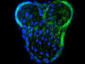
Cell Attachment Bioassay
To demonstrate cell attachment, cells were seeded onto the SphereCol®, human collagen coated bead surface. The photo below is a fluorescent image of human Mesenchymal Stem Cells (HMSC) on SphereCol® beads. DAPI stained nuclei appear blue and phalloidin-Alexa-488 staining of actin filaments is green.